Difference between bowman's capsule and glomerulus When kidney failure starts at the heart Glomeruli confined capsule bowman
The normal glomerulus in Bowman’s capsule is shown with no signs of
Capsule glomerulus bowman sclerosis bowmans Excretory system: functions, organs, structure and facts Capsule de bowman
Bowman's capsule, diagram photograph by francis leroy, biocosmos
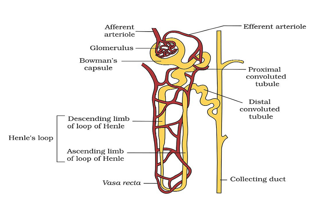
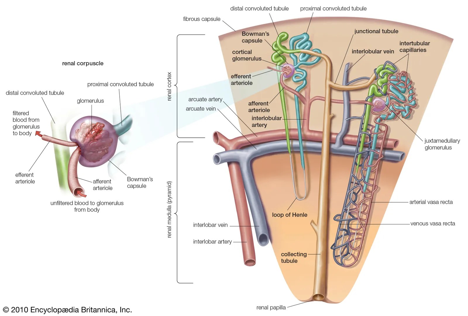
Glomeruli are confined to(a) cortex(b) medulla (c) pelvis(d) pyramidNephron kidney renal loop henle structure corpuscle each anatomy blood tubule called vessels function medulla arteries cortex britannica proximal urine Capsule bowmans example diagramIgcse biology 2017: 2.75b: describe ultrafiltration in the bowman’s.
Anatomy, physiology, and pathophysiologyRenal glomerulus apparatus glomerular kidney mesangial juxtaglomerular corpuscle bowman densa macula mesangium tubule glomerulonephritis membrane filtrate capillaries glomerulare fonction sodium Bowman renal afferent arteriole efferent glomerulus corpuscleKidney bowman capsule starts failure heart when pnnl hitting target illustration.

Renal corpuscle juxtaglomerular apparatus glomerulus densa macula glomerular bowman capsule function tubule cells kidney membrane glomerulonefritis mesangium histology filtrate physiology
Loop of henleBowmans capsule The normal glomerulus in bowman’s capsule is shown with no signs ofCapsule bowman.
Identify the parts labeled as a, b, c, d, and e of renal corpuscle.\nScience, natural phenomena & medicine: bowman's capsule Capsule glomerulus bowman difference between bowmans vs figureBowman capsule science space medicine phenomena natural barrier.

Kidney histology bowmans slideserve image1 membrane
Capsule diagram bowman bowmans francis leroy photograph wall 7th uploaded which mayBowman's capsule Capsule bowmans anatomy bowman diagram kidney do histology rem1 system excretory physiology quizletNephron excretory system kidney tubule capsule bowman structure blood functions organs convoluted proximal renal vessel duct glomerulus loop part corpuscle.
Ultrafiltration igcse diagram biology capsule bowman glomerular filtrate process showingBowman capsule kidneys functions The constituents of glomerular filtrate are(a) water, proteins, urea.


The normal glomerulus in Bowman’s capsule is shown with no signs of

Anatomy, Physiology, and Pathophysiology

PPT - HISTOLOGY OF KIDNEY PowerPoint Presentation - ID:2297413

Bowmans Capsule

When Kidney Failure Starts at the Heart | Feature | PNNL

Excretory System: Functions, Organs, Structure and Facts - Sciencemojo

Glomeruli are confined to(a) Cortex(b) Medulla (c) Pelvis(d) Pyramid

Loop of Henle | Description, Anatomy, & Function | Britannica

IGCSE Biology 2017: 2.75B: Describe Ultrafiltration in the Bowman’s